Medical Imaging
Interventional neuro-radiology
In interventional neuro-radiology, therapeutic tools are inserted within the arteries, up to the lesion through a catheter under the control of various image modalities: 2DSA (digital subtracted angiography), 3DRA (3D rotational angiography), MRI. Our team focuses on the use of multimodal systems to help therapeutic interventions. Effective clinical applications are developed in collaborations with the Hospital of Nancy and bore on the segmentation of arterioveinous malformations and the concept of augmented fluoroscopy to help surgeon's gesture.
|
|
Realistic patient-based simulators for planning the embolization of intracranial aneurisms
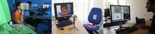
A simulation tool of the interventional act, adapted to the patient's anatomy and physiology, would help to plan the coil placement, train the surgeon, and improve the medical training to the technique. Research on this subject are conducted in collaboration with the Alcove, now SHACRA INRIA group. Our goal is to provide in-vivo models of the patient's organs, and in particular a precise geometric model of the arterial wall with the aim to use these models for real time simulation.
|
|
Augmented reality for deformable organs
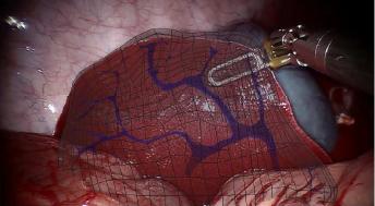
This work is a collaboration with the Shacra team (Lille, Strabourg). We aim at augmenting in real time laparoscopic views in the context of hepatic surgery. Tracking of the liver is done thanks to a bio-mechanical model of the liver guided by image features extracted and tracked on the video at the surface of the organ. Specific models which best suit the considered organs, such as a vascularized model of the liver, have been introduced in this framework. Experiments show that the in-depth localization errors is less than 6mm, and thus below the safety margin required by surgery.
|
|
Image-Driven simulation and the IDeaS ANR project
|
IDeaS is a fundamental research program targeted at per-operative guidance for interventional radiology procedures. Our main goal is to provide effective solutions for the two main drawbacks of interventional radiology procedures, namely: reduce radiation exposure and provide a fully 3D and interactive visual feedback during the procedure. To do so, our project relies on an original combination of computer vision algorithms and interactive physics-based medical simulation. Computer vision algorithms extract relevant information (like the actual projected shape of the guide-wire at any given time) from X-ray images, allowing adjusting the simulation to real data. Conversely, computer-based simulation is used as a sophisticated and trustful predictor for an improved initialization of computer vision tracking algorithms. |


copyright INRIA / Photos C. Lebedinsky